by: Ankita Chatterjee
Shortness of breath and the terrible feeling of choking. An overwhelming fear of nearby danger. An uncontrollable heartbeat. Sudden nausea and dizziness. Any of these symptoms, with no previous warning. These are the ways sufferers of anxiety disorders describe the experience of having an anxiety attack.
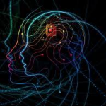
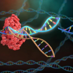
Although current treatments for anxiety do exist, these treatments usually include combinations of medication and therapy. Medications for anxiety disorders generally serve to merely cover up the symptoms of anxiety, and do not target the true molecular basis of the disorder. However, recent research by investigators at UCSF has indicated the existence of specialized neurons within unique regions of the brain that appear to be a physical source of anxiety. Not only have these cells been identified; researchers have also learned to manipulate them in ways that could mitigate the effects of anxiety on the molecular and physical level. This research has fascinating new implications that may one day prove helpful to those suffering from anxiety disorders.
Anxiety disorders have been considered broadly genetic in origin by previous researchers. They have highly complex mechanisms and influencers that aren’t easy to control or understand. The molecular mechanisms of how anxiety is activated, or what parts of the brain “anxiety cells” originate in were previously unknown, but certain sections of the brain such as the hippocampus and hypothalamus appeared to be potential sources for anxiety disorders.
The hippocampus is an essential part of the brain in all stages of life, especially for aspects such as memory, contextual information, navigation, and episodic memory. Additionally, the hypothalamus presented another clue as to the source of anxiety, due to its important role in controlling hormones that influence emotions. Previous research at the University of Texas at Houston linked various different mood disorders to the hippocampus and hypothalamus in the past, but a connection between anxiety and the hippocampal and hypothalamic regions has never been explored on a deeper, cellular level.
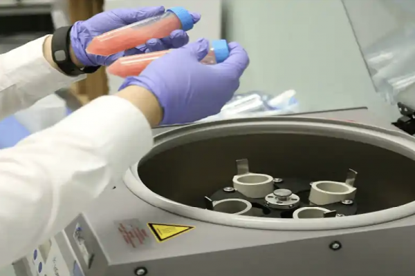
To identify where anxiety originates from within the brain, researchers at UCSF studied the hippocampal activities of mice. “We picked mice because they exhibit a lot of the same behaviors that humans do- when they’re anxious, they tend to not explore their environments,” Dr. Mazen Kheirbek says. Researchers at the Kheirbek Lab used microendoscopic calcium imaging, a kind of imaging that involves miniature microscopes passing light through deep brain tissue, enabling the researchers to monitor the hippocampal and hypothalamic activity of mice. Then, the mice were studied in environments that were known to cause stress and anxiety in rodents, including elevated mazes, platforms, and wide open areas.
The anxiety-related behaviour of the mice was measured by how much they avoided the entrance to the maze or open area, and how much they “head-nodded,” a behavior that signifies anxiousness. Using calcium imaging, the researchers observed that neuronal activity in a part of the hippocampus called the ventral CA1 (vCA1) dramatically increased in mice who were in stressful environments and physically displaying the symptoms of anxiety. Furthermore, the activity of the neurons in this region were directly correlated with the level of anxiety the mice exhibited.
Researchers tested various methods to influence the ways the neurons activated, actually changing the mice’s outward behavior. Optogenetics techniques— implanting mice with fiber optics and illuminating brain tissues with a certain kind of light— effectively “silenced” these neurons. This appeared to control the physical effects of anxiety in the mice, further showing the connection between these neurons and anxiety disorder symptoms. Importantly, these neurons are also present in the same areas of human brains, suggesting a myriad of potential avenues for new anxiety treatments.
Because these neurons and cells are similarly active in the human hypothalamus, further study of this particular region of the brain could lead researchers to a new and more effective anxiety disorder treatment. The vCA1 region has yet to be extensively studied in humans to verify if human anxiety disorders can be controlled in the same way as mouse anxiety. The Kheirbek lab is already looking towards future research to further explore their fascinating findings, and translate their research to the human brain.
“The human hippocampus is larger and a bit more complicated, but in general, it has the same subregions and similar connectivity [as the mouse hippocampus]. What we’re now trying to do is study the genetic profiles of these neurons, and see what makes these cells special,” Dr. Kheirbek says “We can now begin to think about developing non-invasive therapeutics that specifically target these cell types.”
Dr. Kheirbek’s research may be very impactful in the future. One in five adults in the US experiences some kind of anxiety disorder; these disorders and their debilitating effects impact many people in significant and real ways. A truly effective treatment that treats anxiety at its molecular origins would finally provide meaningful relief to sufferers of these disorders, rather than simply masking or altering their symptoms.
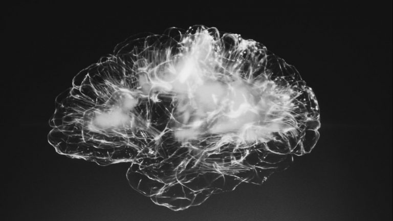
Photo by Alina Grubnyak on Unsplash